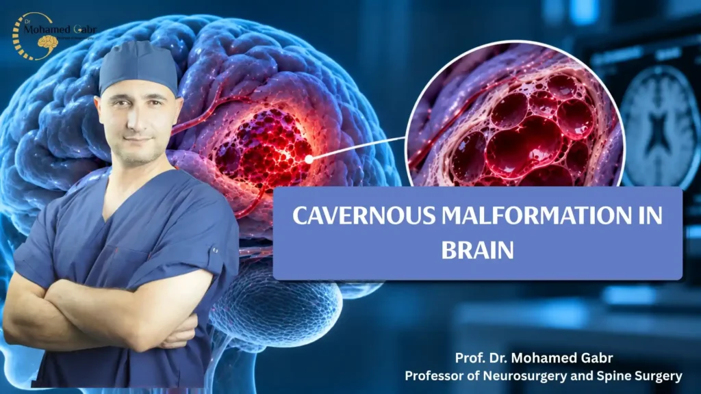
A cavernous malformation in brain is a cluster of abnormal blood vessels that may cause bleeding, seizures, or neurological symptoms.
Although these lesions are noncancerous, their clinical significance depends on location, previous hemorrhage, symptoms, and surrounding neurological structures.
Understanding diagnosis, recurrent bleeding, and treatment options helps patients make informed decisions with qualified neurosurgical specialists.
What Is a Cavernous Malformation in Brain?
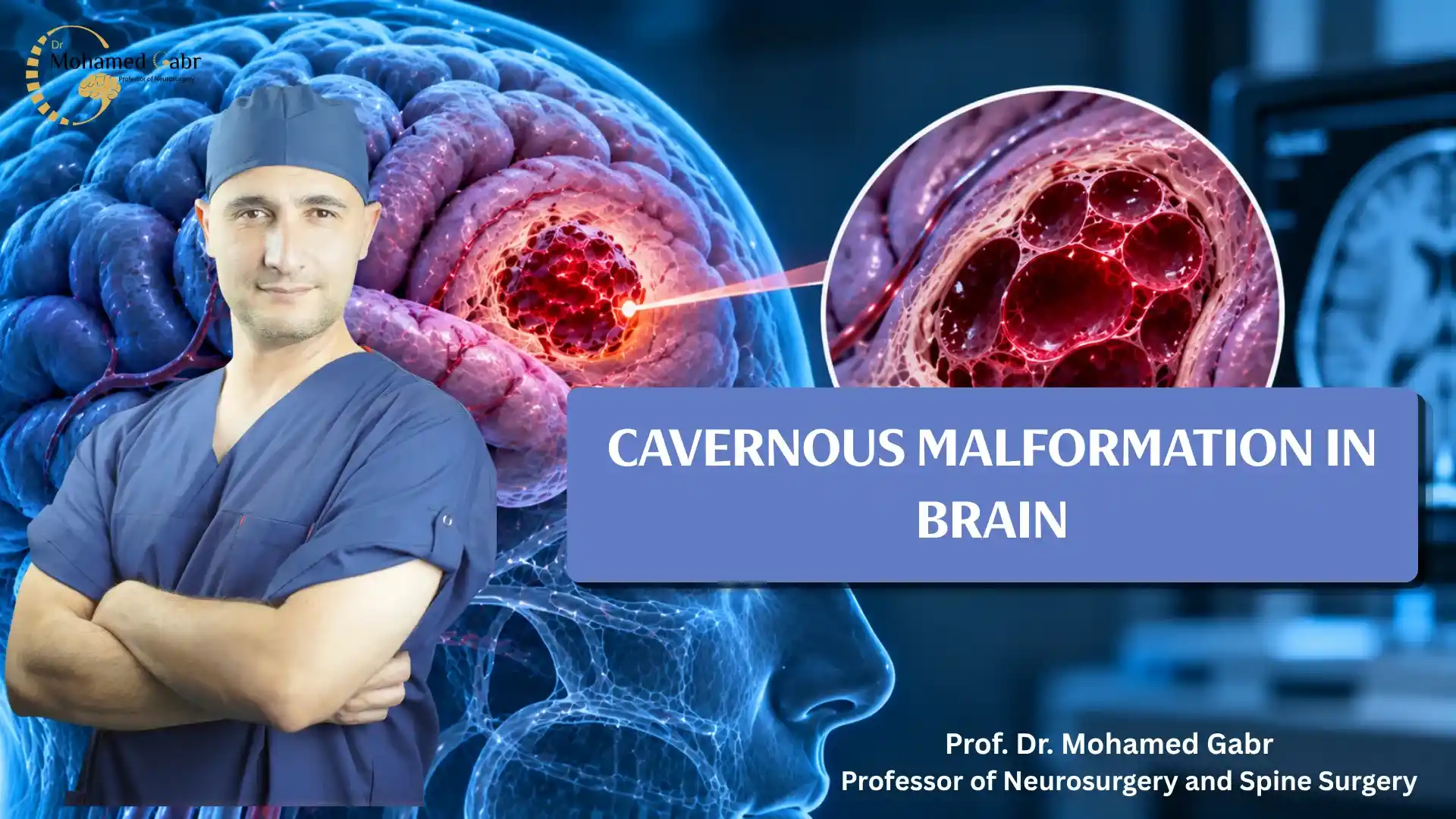
Cerebral cavernous malformations are clusters of abnormal, thin-walled blood vessels characterized by slow blood flow and irregular vascular spaces.
They may develop in different brain regions, including the brainstem, or occasionally within the spinal cord.
Some remain asymptomatic throughout life, whereas others cause seizures, neurological deficits, or symptomatic bleeding affecting nearby brain tissue.
Magnetic resonance imaging (MRI) helps identify these lesions and detect blood products associated with previous hemorrhages.
Causes of a Cavernous Malformation in Brain: Sporadic vs. Genetic (Familial) Cases
Cavernous malformations develop sporadically in many patients, while others inherit genetic variants associated with familial cerebral cavernous malformation disease.
Familial cases commonly involve pathogenic variants in the KRIT1, CCM2, or PDCD10 genes, potentially producing multiple lesions throughout the brain.
A family history or multiple MRI-detected lesions may prompt genetic counselling and testing when clinically appropriate.
Rarely, cavernous malformations develop following brain radiation, so previous radiotherapy is relevant when assessing possible causes.
| Feature | Sporadic cases | Familial cases |
| Cause | No identified familial cause in many cases | Inherited pathogenic genetic variants |
| Lesions | Often solitary; sometimes multiple | Multiple lesions commonly occur |
| Genetics | No familial mutation necessarily identified | KRIT1, CCM2, or PDCD10 variants |
| Family history | May be absent | May include affected relatives |
| Assessment | MRI and clinical evaluation | MRI, family history, possible genetic testing |
The Symptoms of a Cavernous Malformation in Brain
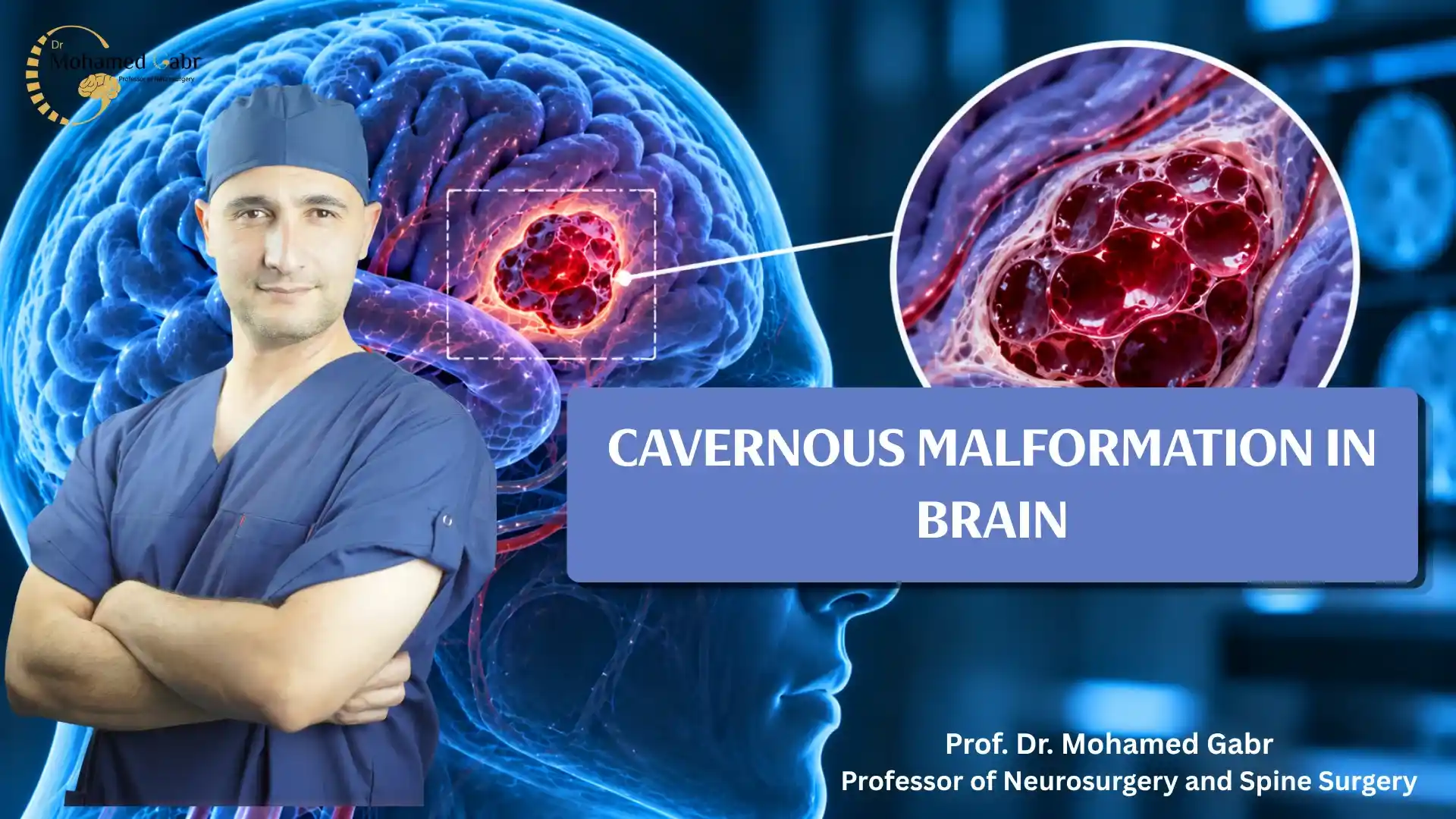
Symptoms depend on lesion location, previous bleeding, and whether nearby neurological pathways become irritated or damaged.
Some patients experience no symptoms, while others develop seizures, headaches, weakness, sensory changes, or coordination difficulties.
Seizures and Epilepsy
Seizures are a common presenting symptom in patients with symptomatic cerebral cavernous malformations, particularly when lesions affect nearby brain tissue.
Blood-breakdown products surrounding a cavernous malformation may irritate nearby brain tissue and contribute to abnormal electrical activity.
However, many cerebral cavernous malformations remain asymptomatic, meaning seizures do not develop in every affected patient.
Neurological assessment, MRI findings, and sometimes electroencephalography (EEG) help specialists evaluate seizures and determine appropriate treatment options.
Focal Neurological Deficits, Headaches, and Brainstem Symptoms
Depending on location, cavernous malformations may affect movement, sensation, speech, balance, swallowing, or eye movements.
Brainstem lesions require particular attention because this region contains essential pathways controlling several neurological functions.
Sudden severe headache, new weakness, speech difficulties, altered consciousness, or prolonged seizures require emergency assessment because serious neurological conditions may be responsible.
Are Cavernous Malformations Dangerous?
A cavernous malformation can cause serious complications, although individual risks vary according to symptoms, location, and previous hemorrhage.
Brainstem location and previous symptomatic bleeding are important considerations when specialists evaluate potential future complications.
However, neither characteristic predicts an individual’s outcome with certainty, and some lesions remain stable without requiring surgery.
If I’ve Had One Bleed From a Cavernous Hemangioma, Is It Likely to Bleed Again?
A previous symptomatic hemorrhage is an important risk factor when specialists assess the possibility of future bleeding from a cerebral cavernous malformation.
However, another hemorrhage is not inevitable. The estimated risk varies according to factors such as lesion location, previous bleeding history, and the patient’s clinical presentation.
Brainstem cavernous malformations may carry different hemorrhage risks from lesions in other brain regions, making individualized assessment particularly important.
Specialists consider the patient’s symptoms, MRI findings, and potential treatment-related complications when comparing continued observation with surgical intervention. A previous bleed alone does not mean that surgery is always necessary.
How Is a Cavernous Malformation Diagnosed?
MRI is the preferred imaging method for diagnosing most cerebral cavernous malformations and evaluating evidence of previous bleeding.
Susceptibility-weighted imaging (SWI), or another suitable susceptibility-sensitive sequence, helps reveal blood products and small lesions that conventional sequences may demonstrate less clearly.
Diffusion tensor imaging (DTI) and tractography may assist surgical planning when lesions lie near important white-matter pathways.
These advanced techniques are not necessary for everyone, and specialists select investigations according to individual clinical circumstances.
Read also: acute subdural hematoma time frame
How Is a Cavernous Malformation Treated?
Treatment decisions depend on symptoms, hemorrhage history, lesion location, accessibility, and potential intervention-related complications.
Observation may suit selected asymptomatic patients, whereas others require medication, further investigation, or assessment for possible microsurgical removal.
Antiseizure medication can help control seizures caused by a cavernous malformation in brain, but it does not remove the underlying vascular lesion.
Read also: dural leak symptoms
Active Observation and Seizure Medication
Active observation may be appropriate when symptoms are absent, and the expected risks of surgery outweigh its potential benefits.
Follow-up plans depend on clinical changes, previous bleeding, imaging findings, and the treating specialist’s recommendations.
Antiseizure medication may control seizures, although persistent seizures sometimes warrant specialist evaluation for possible surgical treatment.
Read also: csf nasal leak symptoms
Complete Microsurgical Resection
Microsurgical resection may benefit selected patients experiencing symptomatic hemorrhage, progressive neurological problems, or difficult-to-control seizures.
Operating microscopes and three-dimensional neuronavigation support anatomical visualization and surgical planning, but they cannot eliminate surgical complications.
For selected complex lesions, diffusion tensor imaging (DTI) tractography and intraoperative neurophysiological monitoring may help surgeons assess nearby neurological pathways.
Complete removal may prevent future hemorrhage from the treated lesion, although it does not guarantee seizure freedom or eliminate bleeding risks from other lesions.
Surgeons generally preserve an associated developmental venous anomaly because disrupting it can cause serious venous complications.
Read also: brain haemorrhage therapies
The Outlook for a Cavernous Malformation: Life Expectancy & Recovery After Surgery
Long-term outcomes vary according to lesion location, previous hemorrhage, neurological injury, and the effectiveness of individualized treatment.
Recovery after surgery differs between patients, with some returning to normal activities while others require rehabilitation.
Surgical removal by a Cerebrovascular Surgery specialist does not guarantee seizure freedom, and some patients still require medication or neurological follow-up.
Why Choose Dr. Mohamed Gabr for Brain Cavernous Malformation Surgery?
Treating a cavernous malformation in brain requires careful assessment and specialized surgical expertise, particularly when recurrent bleeding or drug-resistant seizures are concerns.
Prof. Dr. Mohamed Gabr is a Professor of Neurosurgery and Spine Surgery at Kasr Al Ainy Faculty of Medicine, Cairo University.
He graduated with honors, earned his doctorate in neurosurgery, and received specialized training at Barrow Neurological Institute and Augsburg Hospital in Germany.
His professional background includes complex neurological conditions requiring careful vascular assessment and individualized discussions about appropriate treatment options.
- Risk Assessment for Recurrent Bleeding: Reviewing MRI findings, previous hemorrhages, lesion location, and symptoms helps assess the risks of observation and surgical intervention. Treatment recommendations should reflect each patient’s clinical circumstances rather than assuming every cavernous malformation requires surgery.
- Microsurgical Resection When Indicated: Surgical microscopes and three-dimensional neuronavigation may improve anatomical visualization and support operative planning. Whether resection is appropriate depends on lesion location, symptoms, expected benefits, and potential complications. Complete removal may prevent future hemorrhage from the treated lesion but cannot guarantee seizure freedom.
- Planning for Deep and Brainstem Lesions: DTI fiber tractography and intraoperative neurophysiological monitoring may help assess critical neurological pathways during selected procedures. These technologies support surgical planning but cannot eliminate all complications or guarantee preservation of motor, speech, and cranial nerve functions.
FAQ About Cavernous Malformation in Brain
This FAQ about cavernous malformation in brain answers common questions about symptoms, bleeding risks, diagnosis, treatment options, microsurgery, and recovery.
Can cavernous malformations cause seizures?
Yes, cavernous malformations can irritate surrounding brain tissue and contribute to seizures, although many lesions remain asymptomatic throughout life.
Does every cavernous malformation require surgery?
No, observation may be appropriate for selected patients, depending on symptoms, previous bleeding, lesion location, and surgical risks.
Which MRI technique helps diagnose cavernous malformations?
MRI with susceptibility-weighted imaging helps identify cavernous malformations and blood products associated with previous hemorrhage.
Can surgery prevent another hemorrhage?
Complete removal may prevent future hemorrhage from the treated lesion, but surgical complications and bleeding from other lesions remain possible.
Conclusion
A cavernous malformation in brain requires individualized assessment because symptoms, bleeding risks, and appropriate treatment options can vary considerably between patients.
Consult a qualified specialist to discuss imaging findings, monitoring, medication, and whether microsurgical treatment offers meaningful potential benefits.
Seek emergency medical attention for sudden neurological symptoms, including severe headache, weakness, speech difficulties, or prolonged seizures.
Sources
- Akers et al. — Consensus Guidelines for the Diagnosis and Clinical Management of Cavernous Malformations
- Mayo Clinic — Cavernous Malformations: Symptoms and Causes
- Mayo Clinic — Cavernous Malformations: Diagnosis and Treatment
- Alliance to Cure Cavernous Malformation — CCM Care Guidelines, 2025 Update
- Alliance to Cure Cavernous Malformation — Imaging and Monitoring