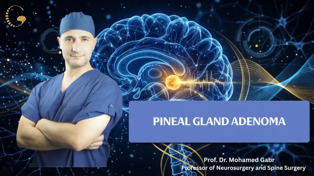
Pineal gland adenoma symptoms and treatment depend on the lesion’s type, size, and effects on nearby brain structures.
However, this term does not describe all pineal region tumors, which include distinct conditions with different growth patterns and treatment needs.
Accurate diagnosis helps doctors determine whether monitoring, surgery, radiation therapy, or other treatments are appropriate for each patient.
What is a Pineal Gland Adenoma and Where is it Located?
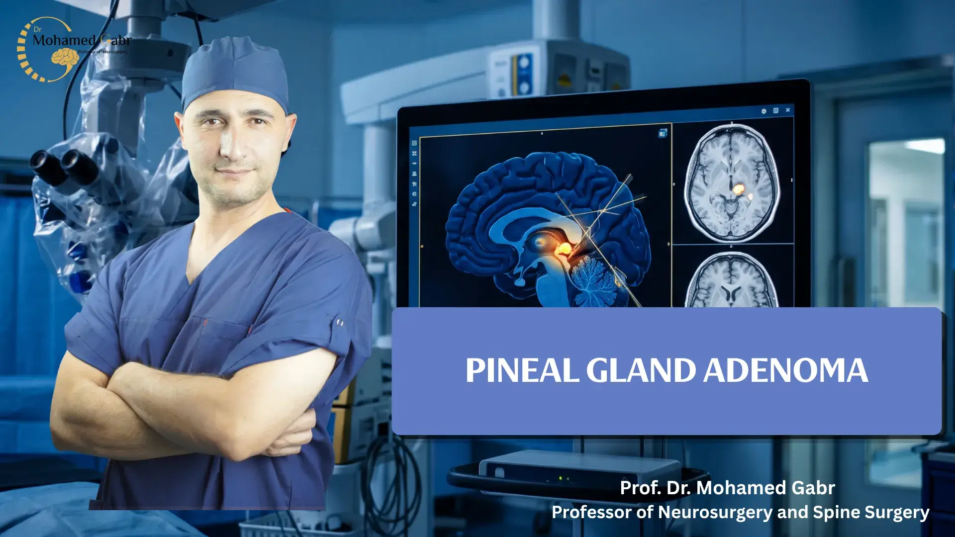
The pineal gland is a small structure deep within the brain that produces melatonin, a hormone involved in regulating the sleep–wake cycle.
A growth near this gland is called a pineal region tumor. These tumors develop from different cell types and vary in behavior.
A tumor near the cerebral aqueduct may block cerebrospinal fluid flow, causing hydrocephalus and increasing pressure inside the skull.
Pineal Cysts vs. Pineal Gland Adenoma: What’s the Difference?
A pineal cyst is a fluid-filled sac, usually benign and often discovered incidentally during brain imaging. Many remain stable without treatment.
Pineal region tumors involve abnormal cell growth. Some grow slowly, while others behave aggressively or spread through cerebrospinal fluid.
MRI helps distinguish cysts from solid masses and assess nearby structures. Further evaluation depends on imaging findings, symptoms, and suspected diagnosis.
What Causes a Pineal Gland Adenoma?
The exact cause of most pineal region tumors remains unknown, and researchers continue investigating their underlying biological mechanisms.
Certain inherited genetic changes may increase the risk of pineoblastoma, including alterations in the RB1 and DICER1 genes.
These genetic associations apply only to specific cases and do not explain the development of most pineal region tumors.
Types and Grades of Pineal Gland Tumors
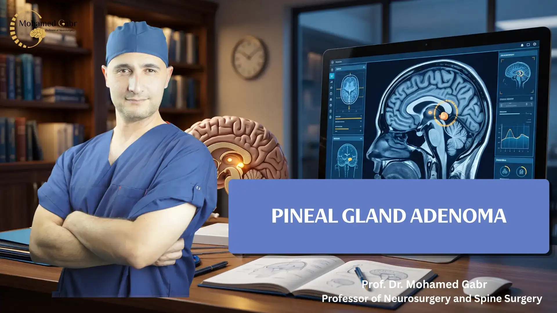
Pineal region tumors include pineal parenchymal tumors, germ cell tumors, and papillary tumors that differ in their biological behavior.
Therefore, accurate classification helps doctors select appropriate treatment options based on each tumor’s type and biological behavior.
Pineocytoma vs. Pineoblastoma (Grades I to IV)
Pineocytoma is generally a slow-growing, grade 1 tumor with relatively favorable biological behavior in many diagnosed cases.
In contrast, PPTID is usually classified as grade 2 or 3, while pineoblastoma is a more aggressive grade 4 tumor.
Importantly, these grades distinguish tumor types according to their biological characteristics rather than describing stages of one disease.
Therefore, treatment decisions depend on the pathological diagnosis, tumor location, and whether the tumor has spread elsewhere.
Papillary Tumor of the Pineal Region (PTPR)
PTPR is a rare tumor that develops in the pineal region but differs from pineal parenchymal tumors in origin and characteristics.
Depending on its pathological features and clinical behavior, surgery may be considered, followed by additional treatment or regular monitoring when appropriate.
What Are the Symptoms of a Pineal Region Tumor?
Symptoms vary according to the tumor’s size, location, growth rate, and effect on cerebrospinal fluid circulation.
However, some lesions cause no symptoms and are discovered incidentally during imaging performed for another medical reason.
When symptoms occur, they may include persistent headaches, nausea, vomiting, blurred or double vision, balance difficulties, and changes in walking.
Symptoms Related to Hydrocephalus (Fluid Buildup in the Brain)
When a tumor blocks cerebrospinal fluid circulation, hydrocephalus may develop and increase pressure inside the skull. (17 كلمة)
As a result, rising pressure can cause symptoms that require prompt medical attention and appropriate clinical evaluation.
These symptoms may include worsening headaches, repeated vomiting, unusual drowsiness, and difficulty walking or maintaining balance during daily activities.
If these symptoms become severe or worsen rapidly, urgent medical assessment is necessary to identify the cause and prevent complications.
Vision and Eye Movement Problems (Parinaud’s Syndrome)
Tumors in the pineal region may affect nearby structures responsible for controlling eye movements and coordinating normal visual function.
Consequently, Parinaud’s syndrome can develop, causing difficulty looking upward and sometimes producing additional visual disturbances.
Because these symptoms can have several underlying causes, neurological assessment and appropriate imaging are needed to identify the problem accurately.
How Is a Pineal Tumor Diagnosed?
Diagnosis begins with a review of the patient’s medical history, a neurological examination, and brain imaging to assess the tumor.
MRI with contrast provides detailed images of the tumor and nearby structures. Meanwhile, CT may reveal calcification or hydrocephalus.
Doctors may recommend additional tests based on the suspected tumor type, imaging findings, and the patient’s clinical condition.
When medically appropriate and safe, a biopsy or surgical tissue sample may help confirm the suspected tumor diagnosis.
However, doctors select further investigations according to symptoms, imaging results, and the patient’s overall clinical condition.
Treatment Options for Pineal Gland Adenoma
Treatment for pineal region tumors depends on the specific diagnosis, tumor size, location, symptoms, and biological behavior.
In particular, doctors consider its type, size, location, symptoms, and evidence of spread when determining the most appropriate treatment.
Depending on the tumor type, size, and location, brain tumor treatment may involve surgery, radiation therapy, chemotherapy, or a combination of these approaches tailored to the patient’s condition.
Endoscopic Treatment for Hydrocephalus (ETV)
Endoscopic third ventriculostomy (ETV) creates an opening that allows cerebrospinal fluid to bypass an obstruction in selected patients.
This procedure may relieve hydrocephalus and reduce pressure inside the skull for selected patients with obstructed cerebrospinal fluid flow.
However, ETV does not remove the tumor itself, so additional treatment or monitoring may still be necessary.
Read also: acoustic neuromas schwannomas symptoms
Microsurgical Tumor Removal (Keyhole & Navigation-Guided Surgery)
Microsurgery may provide tissue for diagnosis, reduce tumor size, or remove as much of the lesion as safely possible.
A keyhole approach may be appropriate for selected patients, depending on individual anatomy and the tumor’s specific characteristics.
The pineal region lies near important veins and neurological structures. Therefore, neuronavigation helps surgeons use preoperative imaging to plan and guide surgery.
However, surgeons choose the approach according to tumor size, location, and proximity to critical surrounding structures.
Complete removal is not always safe or possible. Therefore, surgeons must balance potential benefits against the risks of damaging nearby structures.
Read also: adenoma of the brain
Radiation Therapy and Radiosurgery
Radiation therapy may be recommended after surgery or for tumors that respond well to this treatment approach.
In addition, chemotherapy may be necessary for selected tumor types, including certain germ cell tumors and pineoblastomas.
Stereotactic radiosurgery delivers focused radiation to target tumors without requiring an open surgical incision or conventional surgical exposure.
However, its suitability depends on the tumor’s diagnosis, size, location, biological behavior, and the patient’s overall treatment plan.
Why Choose Dr. Mohamed Gabr for Complex Pineal Tumor Surgery?
Pineal region tumor surgery is highly complex because the gland lies deep within the brain, near critical veins and the brainstem.
Dr. Mohamed Gabr evaluates each case individually, focusing on selecting the most appropriate surgical approach while preserving neurological function whenever possible.
Minimally Invasive Endoscopic Management: Endoscopic third ventriculostomy (ETV) may help relieve hydrocephalus by improving cerebrospinal fluid circulation in selected patients.
When appropriate and safe, an endoscopic procedure may also allow doctors to obtain a tumor biopsy for accurate pathological diagnosis.
In selected patients, ETV may reduce the need for a permanent shunt, depending on the cause of hydrocephalus and clinical findings.
Microsurgery & 3D Neuronavigation: Surgical microscopes and neuronavigation systems help surgeons plan access to deep-seated pineal region tumors with greater precision.
These technologies support surgical guidance while helping protect nearby blood vessels, critical neurological structures, and healthy brain tissue during surgery.
Accurate Differentiation Between Cysts & Tumors: Careful clinical assessment and brain imaging help distinguish benign pineal cysts from tumors requiring further evaluation.
Pineal cysts often require monitoring, while treatment decisions for tumors depend on diagnosis, lesion characteristics, symptoms, and individual clinical findings.
Conclusion
Pineal gland adenoma symptoms and treatment depend on the tumor’s type, growth pattern, biological behavior, and individual treatment needs.
Therefore, accurate imaging and diagnosis help doctors select the most appropriate management plan for each patient.
Depending on the findings, treatment may involve monitoring, microsurgery, hydrocephalus management, or additional therapies when necessary.
Ultimately, patients should discuss their diagnosis, available treatment options, and potential risks with a qualified specialist.
Sources
- Mayo Clinic: Pineal Gland Tumor — Symptoms and Causes
- Mayo Clinic: Pineal Gland Tumor — Diagnosis and Treatment
- National Cancer Institute: Pineal Region Tumors — Diagnosis and Treatment
- Cedars-Sinai: Pineal Tumor
- Columbia University Department of Neurological Surgery: Pineal Region Tumor
Frequently Asked Questions (FAQ)
This section answers common questions about pineal region tumor symptoms, diagnosis, treatment options, surgical risks, and recovery following treatment.
1. Is a pineal gland adenoma cancerous?
The term does not identify one specific tumor type. Therefore, pineal region lesions may be benign or malignant, making an accurate diagnosis essential.
2. Can a pineal region tumor be removed completely?
Some tumors can be removed completely. However, the possibility depends on the tumor’s type, location, and relationship to critical structures.
3. Does ETV remove a pineal tumor?
No. ETV allows cerebrospinal fluid to bypass an obstruction. Consequently, it may relieve hydrocephalus, but it does not remove the tumor itself.
4. Do all pineal region tumors require surgery?
Instead, treatment depends on the diagnosis. Some tumors require monitoring, surgery, radiation therapy, chemotherapy, or combined treatment.