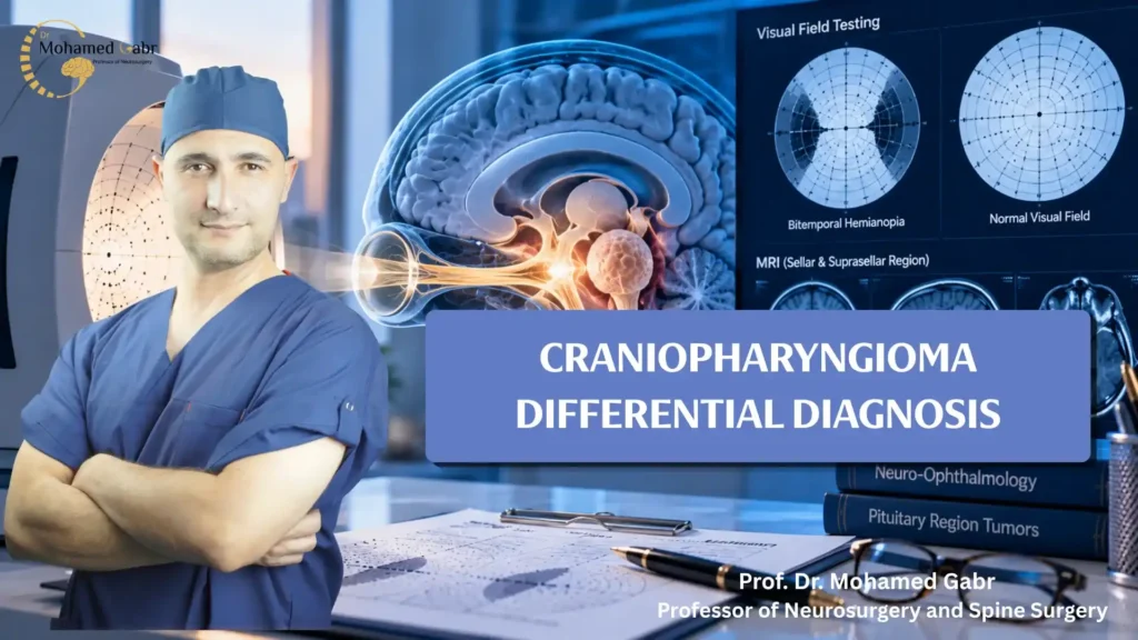
Craniopharyngioma Differential Diagnosis helps doctors distinguish craniopharyngiomas from other lesions affecting the pituitary gland, hypothalamus, and nearby visual pathways.
Although these conditions may look similar on MRI, their origins, risks, and appropriate treatments can differ considerably.
Therefore, accurate diagnosis combines imaging, symptoms, hormonal testing, and specialist assessment before doctors recommend monitoring or surgery.
What Is a Craniopharyngioma?
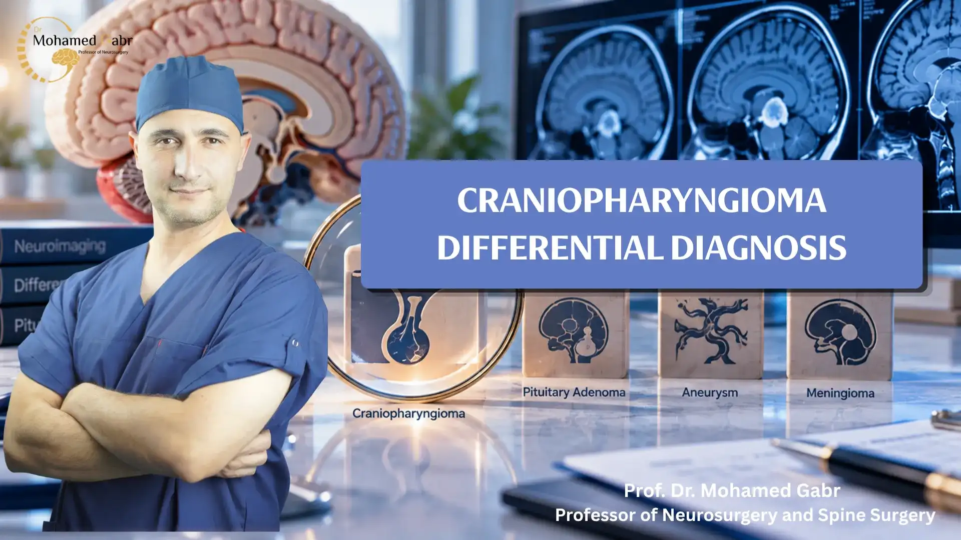
A craniopharyngioma is a typically slow-growing tumor that develops near the pituitary gland and hypothalamus.
Although usually classified as benign, its location can cause serious complications involving vision, hormone production, and neurological function.
For example, pressure on nearby structures may cause headaches, visual disturbances, growth problems, or hormonal deficiencies.
Additionally, these tumors may contain solid tissue, cysts, and calcifications, creating appearances that overlap with other skull base lesions.
Why is a Craniopharyngioma Differential Diagnosis Crucial for Treatment?
Accurate diagnosis matters because similar-looking lesions may require observation, surgery, or specialized treatment rather than identical management.
For example, selected asymptomatic cysts may need monitoring, whereas lesions threatening vision may require more active intervention.
Furthermore, mistaking an aneurysm for a tumor could expose patients to dangerous bleeding during an inappropriate surgical procedure.
Therefore, doctors evaluate imaging, symptoms, hormonal results, and clinical history before determining the safest management strategy.
Common Conditions in Craniopharyngioma Differential Diagnosis
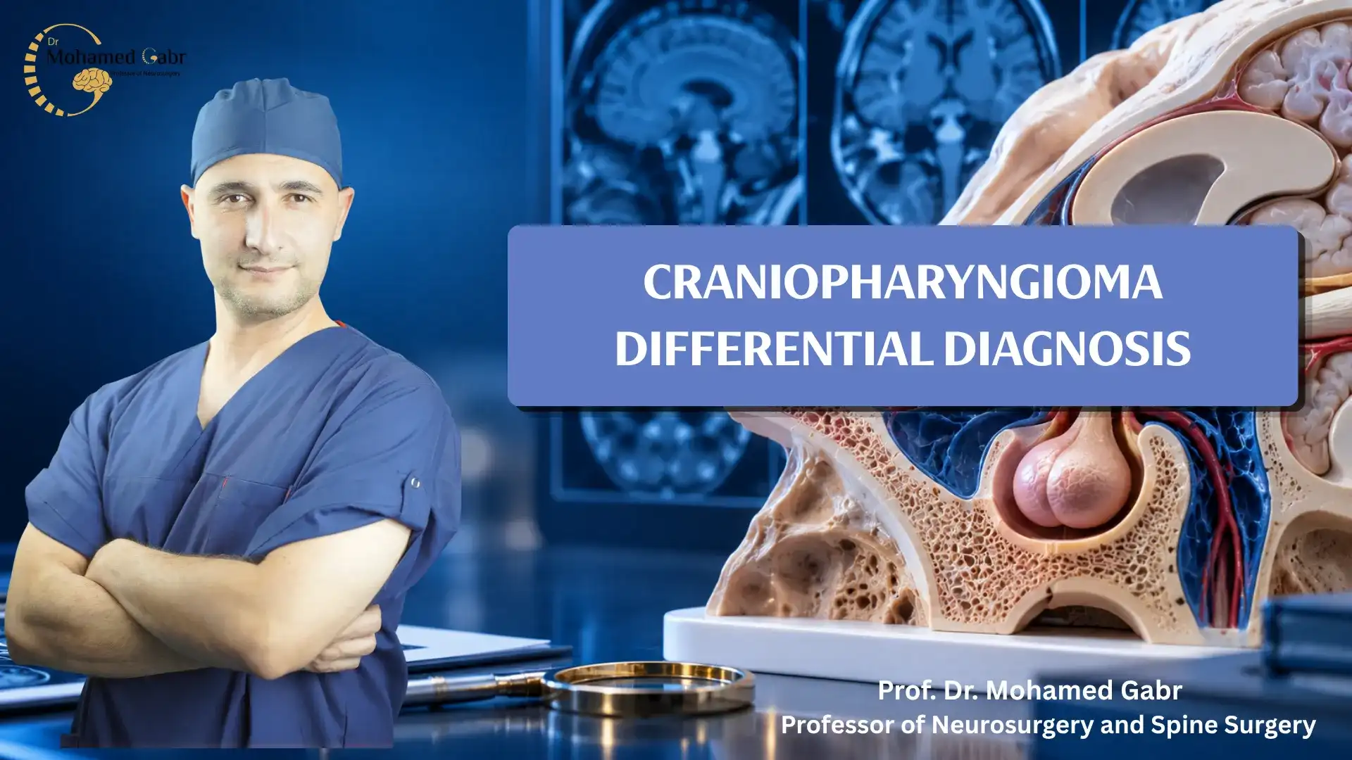
Several lesions resemble craniopharyngiomas because they develop near the pituitary gland or contain fluid-filled spaces.
However, their imaging characteristics, biological behavior, and clinical effects help specialists distinguish these conditions.
Rathke’s Cleft Cysts
Rathke’s cleft cysts are benign lesions arising from embryological remnants near the pituitary gland.
Unlike craniopharyngiomas, these cysts generally lack a solid tumor component, although their appearance varies with fluid composition.
Nevertheless, larger cysts may compress nearby structures, causing headaches, visual disturbances, or hormonal abnormalities.
Therefore, doctors consider cyst location, wall appearance, enhancement patterns, symptoms, and endocrine findings when evaluating these lesions.
Cystic Pituitary Adenomas
Pituitary adenomas, also called pituitary neuroendocrine tumors, develop within the gland and sometimes contain cystic or hemorrhagic components.
Unlike craniopharyngiomas, some adenomas produce excess hormones, although others remain hormonally inactive.
For example, prolactin testing may reveal abnormalities supporting a pituitary tumor diagnosis rather than another skull base lesion.
However, imaging appearances can overlap, especially when larger adenomas extend above the gland or undergo internal degeneration.
Consequently, specialists assess tumor origin, enhancement, hormone production, and relationships with surrounding anatomical structures.
Meningiomas and Optic Pathway Gliomas
Meningiomas arise from the membranes surrounding the brain and may develop near the optic nerves or pituitary region.
Typically, these tumors demonstrate solid enhancement on MRI, helping distinguish them from predominantly cystic lesions.
Meanwhile, optic pathway gliomas affect visual pathways and may cause progressive visual impairment, particularly among children.
Although their locations can resemble craniopharyngiomas, their tissue characteristics and growth patterns often differ.
Therefore, radiologists examine lesion origin, enhancement, and relationships with nearby nerves and other structures.
Epidermoid and Dermoid Cysts
Epidermoid and dermoid cysts are developmental lesions that occasionally occur near skull base structures and resemble other masses.
However, diffusion-weighted MRI often helps identify epidermoid cysts because they characteristically demonstrate restricted diffusion.
By comparison, dermoid cysts may contain fat, producing distinctive signals on MRI and appearances on computed tomography.
Nevertheless, findings vary according to cyst contents and location, so specialists interpret multiple imaging sequences together.
Skull Base Aneurysms
A skull base aneurysm is an abnormal blood vessel dilation that may resemble a mass near the pituitary region.
Unlike tumors, aneurysms involve blood vessels, making accurate identification essential before considering invasive treatment.
Therefore, doctors may request CT angiography or MR angiography when initial imaging raises vascular concerns.
Importantly, recognizing an aneurysm before surgery helps prevent accidental vessel injury and potentially life-threatening bleeding.
How Do Neurosurgeons Differentiate These Conditions?
MRI helps doctors assess soft tissues, cyst contents, tumor enhancement, and relationships with nearby neurological structures.
Additionally, contrast-enhanced sequences may reveal solid tissue or cyst wall enhancement, providing useful diagnostic clues.
Meanwhile, diffusion-weighted imaging helps identify epidermoid cysts, while CT can demonstrate calcifications and skull base bone changes.
If vascular involvement remains possible, angiographic imaging clarifies relationships between the lesion and nearby arteries.
Nevertheless, imaging cannot always establish a definitive diagnosis, so doctors also consider symptoms and hormonal test results.
In selected cases, pathological examination provides additional evidence to confirm the lesion’s nature.
Read also:
Symptoms Shared by Craniopharyngiomas and Similar Skull Base Lesions
Because these lesions develop in nearby anatomical regions, they may produce similar symptoms despite having different causes.
Therefore, symptoms alone rarely establish the diagnosis, making clinical examination and appropriate investigations essential.
Read also: brain tumor symptoms in adults
Vision Loss and Visual Field Defects
Compression of the optic nerves or chiasm may cause blurred vision, reduced peripheral vision, or other visual field defects.
However, symptoms vary according to lesion location, size, growth, and pressure on surrounding visual pathways.
Therefore, formal visual field testing helps document impairment and monitor changes during treatment.
Hormonal Imbalances and Growth Issues
Lesions near the pituitary gland may disrupt hormone production, causing fatigue, menstrual changes, or growth abnormalities.
Additionally, hypothalamic involvement may disturb water balance, sometimes causing excessive thirst and frequent urination.
However, these symptoms also occur with other endocrine disorders, so doctors investigate their underlying causes.
Consequently, blood tests, urine studies, and specialist assessments help identify hormone deficiencies or excessive hormone production.
Read also: benign brain tumor symptoms in women
Advanced Surgical Treatment Options for Skull Base Tumors & Cysts
Treatment depends on the confirmed diagnosis, lesion size, symptoms, growth, and risks to surrounding structures.
For example, selected asymptomatic lesions may undergo monitoring, whereas progressive visual impairment may justify surgical evaluation.
Importantly, not every cyst or tumor requires surgery, because management must reflect individual clinical findings.
Read also: brain tumor stages and symptoms
Minimally Invasive Endoscopic Endonasal Surgery
Endoscopic endonasal surgery allows selected skull base lesions to be reached through the nasal passages using specialized instruments.
In suitable cases, this approach avoids an external incision and provides access to lesions near the pituitary gland.
However, suitability depends on lesion position, vascular relationships, extension, and the surgical team’s experience.
Furthermore, potential complications include cerebrospinal fluid leakage, hormonal disturbances, infection, and injury to nearby structures.
Precision Microsurgery via Keyhole Approaches
For selected lesions, microsurgical or keyhole approaches provide access through carefully planned openings while limiting tissue disruption.
Nevertheless, the safest approach depends on lesion anatomy, vascular involvement, extension, and proximity to critical structures.
Therefore, neurosurgeons compare available techniques to balance lesion treatment with preservation of vision and neurological function.
Read also: chromophobe adenoma symptoms
Why Choose Dr. Mohamed Gabr for Complex Skull Base Diagnosis and Surgery?
Accurately diagnosing skull base lesions, including craniopharyngiomas and their mimics, requires specialized expertise because they lie near critical nerves and major arteries.
Dr. Mohamed Gabr brings extensive expertise in complex skull base and pituitary neurosurgery, ensuring diagnostic precision and surgical safety through:
- Advanced Radiological Analysis: Meticulous evaluation of specialized MRI sequences to accurately differentiate true neoplasms from benign cysts (like Rathke’s cleft cysts), preventing inappropriate or unnecessary surgical interventions.
- Scarless Endoscopic Endonasal Approach: Utilizing state-of-the-art neuro-endoscopy to access deep skull base lesions through the natural nasal passages, achieving complete tumor or cyst resection without external incisions or head shaving.
- Optic Nerve Protection & Vision Restoration: Employing advanced 3D Neuronavigation and surgical microscopes to safely decompress the optic chiasm, safeguarding and effectively restoring the patient’s vision post-surgery.
FAQ
Below are answers to common questions about craniopharyngioma diagnosis, similar skull base lesions, imaging methods, symptoms, and treatment options.
Can MRI always distinguish craniopharyngiomas from similar cysts?
MRI often provides useful diagnostic clues. However, some lesions overlap in appearance and require additional imaging or specialist interpretation.
Can Rathke’s cleft cysts affect vision?
Yes. In particular, larger cysts can compress nearby visual pathways or the pituitary gland, potentially causing visual disturbances or hormonal symptoms.
Does every craniopharyngioma require surgery?
No. Instead, management depends on symptoms, growth, lesion characteristics, and surrounding anatomy; specialists determine the most appropriate treatment plan.
Why must doctors rule out an aneurysm?
An aneurysm involves a blood vessel. Therefore, invasive treatment without recognizing it may cause dangerous bleeding or other serious complications.
Conclusion
An accurate Craniopharyngioma Differential Diagnosis helps distinguish tumors, benign cysts, and vascular abnormalities that may appear similar.
Although MRI provides essential information, doctors must also consider symptoms, hormonal results, anatomy, and possible vascular involvement.
Ultimately, individualized assessment helps specialists select appropriate management while prioritizing vision, endocrine function, and patient safety.
References
- https://www.ncbi.nlm.nih.gov/books/NBK538819/
- https://doi.org/10.1016/j.ando.2024.07.002
- https://doi.org/10.1148/rg.2021200203
- https://doi.org/10.3390/cancers16142532
- https://doi.org/10.3389/fendo.2025.1630695